Abstract
Classical clinico-anatomical correlations traditionally linked symptoms to specific lesions and local deficits. However, in recent decades, research from lesion studies, functional MRI (fMRI), and white-matter neuroanatomy has increasingly defined many clinical phenomena as failures of large-scale network function. This article explores the evolution from early concepts of diaschisis and disconnexion to modern network phenomenology. It examines how fMRI, lesion-network mapping, and tract-based neuroanatomy support and limit network explanations of cognition, behavior, and consciousness. The article discusses translational implications for prognosis and neuromodulation, arguing that the most significant progress occurs when functional network models are anchored to testable structural pathways and causal perturbations, with explicit acknowledgment of methodological limitations.
Introduction
A century ago, von Monakow introduced the concept of diaschisis, describing remote functional depression following focal injury, suggesting that symptoms could emerge from disrupted interactions rather than tissue loss alone. Mid-20th-century neuropsychology expanded this dynamic systems view, culminating in Geschwind's "disconnexion syndromes," which emphasized the role of white matter and interareal communication in human neurology. These historical shifts laid the foundation for today's network neuroscience, where clinical syndromes are increasingly understood as emergent properties of disrupted large-scale communication rather than isolated nodal failures.
From Modules to Networks
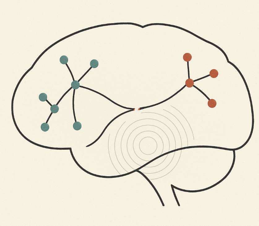
The modern network era began when intrinsic activity patterns, especially the default mode network (DMN), revealed a structured baseline architecture that is dynamically suspended during external task engagement. Subsequent research identified at least three interacting systems: a frontoparietal "executive" network (FPN), a salience network (SN) anchored in the anterior insula/dorsal anterior cingulate, and the DMN. The right fronto-insular cortex plays a crucial role in switching between the DMN and FPN, linking interoception and control to network state transitions. These findings shifted the focus from local activation maps to inter-network coordination and its breakdown in disease.
Simultaneously, structure-function studies demonstrated that functional connectivity is constrained, though not determined, by anatomical coupling. The macroscopic scaffold, comprising long-range association tracts and hub "rich-club" architecture, supports integration across modules. Damage to hubs disproportionately degrades multiscale communication and highlights transdiagnostic vulnerability. These insights motivate a mechanistic perspective: clinical symptoms often reflect losses of network controllability and communication capacity rather than focal neuronal failure.
fMRI Evidence for Network Phenomenology
Resting-state and task fMRI consistently reveal distinct but interacting networks that support internal mentation (DMN), cognitive control (FPN), and salience-driven reorientation (SN). Aberrant SN-mediated access/engagement of DMN and FPN has been proposed as a unifying explanation for diverse psychiatric and neurological disorders, reframing heterogeneous symptom clusters as network-level dysmetria. Causal and chronometric analyses identify the right fronto-insula as critical for rapid switches between internally and externally oriented modes, suggesting that clinical "fluctuations" (e.g., lapses in awareness or goal maintenance) are failures of network state transitions.
However, network inferences from BOLD signals come with caveats: motion confounds, preprocessing choices (e.g., global-signal regression), and inflated cluster-wise false positives can mislead. This highlights the need to corroborate fMRI findings with convergent methods before establishing causality.
Fascicular (Tract-Based) Anatomy: From Association Streams to Causal Pathways
Ventral and dorsal "streams" in language extend to specific long association bundles with distinct cognitive functions. Direct stimulation and lesion studies implicate the inferior fronto-occipital fasciculus (IFOF) in semantic control and integration; disruption leads to semantic paraphasia or non-verbal semantic deficits. The frontal aslant tract (FAT), connecting inferior frontal regions with pre-SMA/SMA, contributes to speech initiation/fluency and broader sequential planning. Branches of the superior longitudinal fasciculi (SLF) support spatial attention; right-hemisphere SLF II/III disconnection predicts neglect severity. These fascicles form the structural backbone of networks revealed by fMRI and provide causal leverage in clinical settings.
Nevertheless, diffusion-tractography has significant error modes (false positives/negatives, gyral bias, crossing fibers). Ground-truth challenges show wide variability across algorithms, so fascicular claims must be verified with intraoperative mapping, lesion-symptom analyses, and cross-modal replication.
Lesions, Networks, and Causality
Traditional clinico-radiologic correlation explains deficits when lesions overlap a necessary node; however, many neuropsychiatric phenomena arise when distinct lesion sites converge on a common network. Lesion-network mapping (LNM) addresses this by seeding heterogeneous lesion locations into a normative connectome to identify shared connectivity that best predicts the symptom. LNM has localized circuits for depression, criminal behavior, epilepsy, parkinsonism, and amnesia, often implicating distributed networks rather than single loci, thereby reconciling phenotypic similarity with anatomical heterogeneity.
Importantly, LNM is not a cure-all: it inherits limitations of the underlying connectome, is sensitive to lesion sampling, and requires independent validation through stimulation or prospective prediction. Nonetheless, alignment between lesion-derived circuits and effective neuromodulation targets (e.g., left DLPFC sites anti-correlated with sgACC in depression) illustrates how network localization can refine where to intervene when nodal localization fails.
Consciousness as Network Phenomenology
Disorders of consciousness (DoC) highlight the distinction between arousal mechanisms and conscious access, both of which depend on large-scale connectivity. Effective connectivity within lateral frontoparietal circuits differentiates and predicts DoC state, while DMN integrity, though variably impaired, remains a reproducible correlate of residual awareness in group studies. Under anesthesia, transitions in conscious state parallel disruptions and recoveries of frontoparietal-salience interactions, consistent with workspace-style ignition models.
Complementing network topology with perturbational assays, the Perturbational Complexity Index (PCI) - a TMS-EEG measure of the spatiotemporal richness of cortical responses - discriminates levels of consciousness across sleep, anesthesia, and DoC without relying on behavior. PCI operationalizes the idea that consciousness requires both integration and differentiation, providing an orthogonal validation of network-level accounts.
Neurodegeneration and Selective Network Vulnerability
Network models extend beyond acute lesions. In vivo atrophy patterns across distinct neurodegenerative syndromes respect the boundaries of healthy intrinsic networks; the salience network is targeted in behavioral-variant frontotemporal dementia, while default-mode hubs show early functional disruption in amyloid-positive aging and Alzheimer's disease. Such selectivity supports hypotheses of network-mediated spread or shared vulnerability, linking molecular pathology to systems-level topology.
Synthesis: What Makes a Network Account Clinically Useful?
First, multimodal convergence: the most credible network explanations triangulate fMRI dynamics, tract-based anatomy, and causal perturbation (lesion or stimulation). Second, granularity: moving beyond "dual-stream" shorthand to specify which fascicle (e.g., IFOF vs. ILF vs. FAT) carries which computation yields testable predictions and safer surgery. Third, causal validation: LNM and patient-specific connectivity can nominate stimulation targets, but efficacy hinges on prospective, individualized testing. Fourth, methodological humility: both fMRI and tractography have known pitfalls; results should be framed probabilistically and replicated across pipelines and datasets.
Limitations and Open Questions
Network phenomenology risks overextension when functional associations are reified as mechanisms. Structure-function coupling is strong but incomplete; indirect communication, neuromodulatory tone, and time-varying dynamics can dissociate BOLD correlations from axonal pathways. Moreover, rich-club and connector-hub concepts predict vulnerability at scale, yet clinical translation requires patient-specific modeling that integrates lesion burden, tract integrity, and ongoing plasticity. Finally, consciousness research must reconcile workspace-style ignition with posterior "hot-zone" contributions and subcortical arousal systems; perturbational metrics like PCI help, but do not yet map one-to-one onto tractable interventions.
Conclusion
From diaschisis and disconnexion to triple-network dynamics, lesion-network mapping, and fascicular microanatomy, evidence supports a network-centric neurology: many clinical symptoms are better conceived as failures of communication than as mere local deficits. The practical path forward is not to abandon localization but to extend it - localizing computations to edges and pathways, validating models with causal perturbation, and translating maps into individualized, testable interventions.
References
- von Monakow C. Diaschisis: origins and modern perspectives. (Historical/overview).
- Geschwind N. Disconnexion syndromes in animals and man. Brain. 1965.
- Luria AR. Higher Cortical Functions in Man. (English translation; systems view).
- Raichle ME et al. A default mode of brain function. PNAS. 2001.
- Seeley WW et al. Dissociable intrinsic connectivity networks for salience processing and executive control. J Neurosci. 2007.
- Right fronto-insular cortex switches between central-executive and default-mode networks. PNAS. 2008.
- Menon V. Large-scale brain networks and psychopathology: a unifying triple-network model. Trends Cogn Sci. 2011; and updates.
- Honey CJ et al. Predicting functional connectivity from structural connectivity. PNAS. 2009.
- van den Heuvel MP & Sporns O. Rich-club organization of the human connectome. J Neurosci. 2011; Crossley et al. Hubs implicated across brain disorders. Brain. 2014.
- Herbet G et al. Right IFOF and non-verbal semantics. Brain Struct Funct. 2017; Dick AS et al. Frontal Aslant Tract in speech. 2018; Lunven M et al. SLF disconnection and neglect. 2015.
- The challenge of mapping the human connectome based on diffusion tractography. Nat Commun. 2017.
- Boes AD et al. Network localization of symptoms from focal lesions. Brain. 2015; Padmanabhan JL et al. A human depression circuit from lesions. Biol Psychiatry. 2019; Darby RR et al. Lesion-network localization of criminal behavior. PNAS. 2018.
- Ihalainen R et al. Lateral frontoparietal effective connectivity and DoC. PLoS ONE. 2024; Li H et al. Functional networks in prolonged DoC (review). 2023; Mashour GA. Anesthesia and the neurobiology of consciousness. 2024.
- Casali AG et al. Perturbational Complexity Index (PCI). Sci Transl Med. 2013.
- Seeley WW et al. Neurodegenerative diseases target large-scale networks. Neuron. 2009; Sperling RA et al. Amyloid deposition and DMN dysfunction. Neuron. 2009.
- Jones DK & Cercignani M. Pitfalls in diffusion MRI analysis. NMR Biomed. 2010. (fMRI caveats also widely reviewed).