Abstract
The cortical homunculus remains a foundational concept in neurology, illustrating how brain territory is allocated based on behavioral importance. Originating from Penfield and Boldrey's 1937 cortical maps, the homunculus highlights the disproportionate representation of functionally critical body parts. Recent evidence suggests that somatotopy is not limited to the cortex but is also present in subcortical structures like the putamen, globus pallidus, subthalamic nucleus, and thalamus. This review synthesizes historical, anatomical, and clinical findings to emphasize the homunculus's significance in guiding surgery and rehabilitation and interpreting neurophysiological and imaging observations. Understanding somatotopic adjacency - how nearby cortical territories support higher-order functions - enables a nuanced and personalized approach to neurological practice.
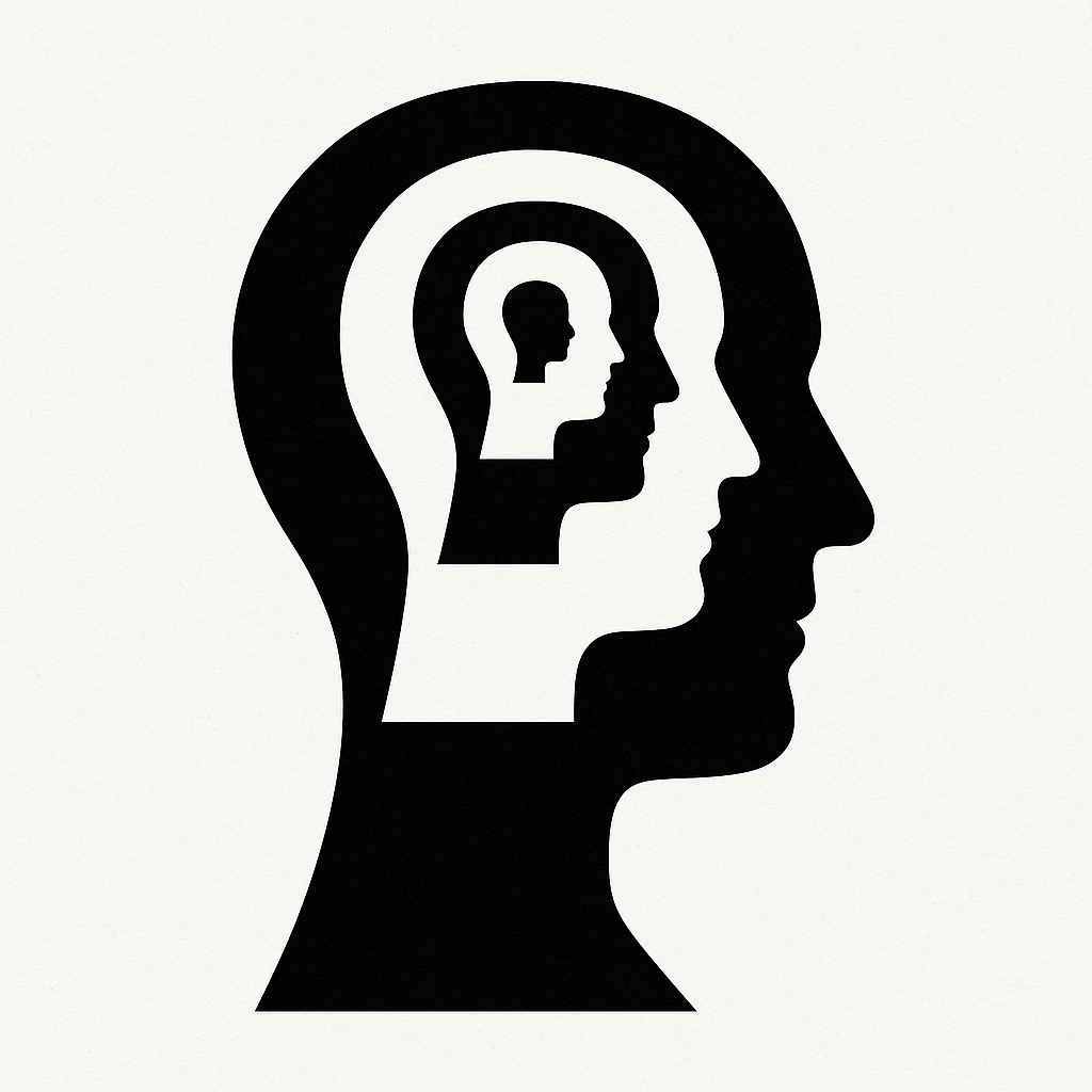
1. Introduction
As a neurologist with a background in neuroscience and evolutionary biology, I view the homunculus as more than a metaphorical shorthand. It is a principle reflecting how neural territory is allocated where behavioral demands are greatest. Penfield's grotesque figure, although imprecise in proportion, underlies gravity in our clinical reasoning. This article revisits the homunculus in light of evolving anatomical knowledge, emphasizing cortical distortions, subcortical preservation, the significance of adjacency, and pragmatic clinical implications even when bench knowledge is incomplete.
2. Historical Foundations
The term homunculus comes from the Latin homo ("man") and the diminutive -culus ("little man"). Wilder Penfield and Edwin Boldrey introduced the cortical motor and sensory maps in 1937, based on electrical stimulation of the human cortex during epilepsy surgery. Their iconic drawings exaggerated the lips, tongue, and hands - body parts with dense representation in cortical maps. The map was refined in later publications, but early diagrams already emphasized cortical distortion based on sensory and motor representation.
Catani et al. (2017) revisited Penfield's original data, analyzing inaccuracies in the depiction. They demonstrated that although the homunculus is not proportional by modern imaging standards, it still reflects systematic mapping of functionally important body parts.
3. Cortical Somatotopy and Functional Significance
The classic somatotopic organization (leg -> trunk -> arm -> hand -> face -> tongue) of M1/S1 reflects the allocation of cortical territory in proportion to behavioral significance and sensory density. Although textbooks present an orderly strip, individual mapping often shows overlap and variation. Nevertheless, the gross layout persists. Functional imaging and direct stimulation continue to demonstrate that regions responsible for fine motor control (hands, face) occupy more cortical space.
4. Subcortical Somatotopy: Preservation Beyond Cortex
Somatotopic organization is preserved in subcortical motor circuits:
- Putamen and GPi
Anterograde and retrograde tracer studies in primates reveal that M1 and SMA projections to the putamen preserve orofacial, forelimb, and hindlimb separations. The GPi also maintains a leg-arm-face gradient. - Subthalamic Nucleus
STN subdivisions show somatotopy via cortico-hyperdirect pathways. Motor territories align with cortical M1/SMA representation patterns. - Thalamic Relay Nuclei
Motor thalamus (e.g., VLo, VApc) preserves topographic mapping consistent with input from the GPi and cerebellum. - Structural Connectivity in GPi
Diffusion MRI connectivity maps confirm somatotopic organization, aiding surgical targeting in DBS.
These preserved subcortical maps are clinically meaningful, allowing selective targeting in DBS, interpretation of movement disorder symptoms, and prediction of lesion outcomes.
5. Adjacency and Functional Clustering
The homunculus teaches that body part representations are adjacent anatomically, but this adjacency underlies functional associations:
- Hand motor zones lie near regions supporting tool use, symbolic manipulation, and praxis.
- Face/mouth motor zones abut language areas (e.g., Broca's area).
- These spatial relationships explain why lesions near the motor cortex may cause deficits in language, tool use, or symbolic tasks that are not confined to motor function.
This "scaffold" allows clinical inference even before bench-validated circuitry is described: lesion location + known adjacency = predictive functional deficit.
6. Clinical Implications
6.1 Surgical Planning
Awake mapping in glioma resection or epilepsy surgery relies on the homuncular framework to preserve function.
6.2 Movement Disorders and DBS
Somatotopy in GPi and STN grounds how we calibrate electrode placement in Parkinson's disease and dystonia.
6.3 Stroke Rehab and Plasticity
Understanding distorted cortical maps guides therapy that harnesses plasticity; functional reallocations can be anticipated and encouraged.
6.4 Interpretive Framework
Even when bench data is incomplete, the homunculus helps interpret clustered symptoms across motor, sensory, and higher functions.
7. Limitations and Ongoing Refinements
- The homunculus is not a precise scale model. Its distortions and overlaps are refined by modern imaging.
- Individual variation matters: mapping must be supplemented by subject-specific data.
- Higher-order functional zones (e.g., math, abstraction) are more distributed and not always adjacent to motor/sensory zones.
Nevertheless, the principle of topographic representation and adjacency remains robust.
8. Conclusion
The homunculus remains a central organizing principle in neurology. Far from being archaic or purely illustrative, it reflects how neural resources are allocated, preserved through cortical and subcortical levels, and organized into functional neighborhoods. As clinicians, we use it not only as a surgical roadmap but also as a guide for interpreting complex symptom clusters, designing rehabilitation strategies, and anticipating potential functional outcomes.
The homunculus reminds us: in the brain's geography, what lies beside matters as much as what lies within - and function follows representation.
References
- Catani M. "A little man of some importance." Brain. 2017;140(11):3055-3061. doi:10.1093/brain/awx270
- Gandhoke GS, et al. Edwin Boldrey and Wilder Penfield's Homunculus: A Life ... World Neurosurgery. 2019;
- Folzenlogen Z, et al. A brief history of cortical localization and its mapping... Neurosurg Focus. 2019;47(3):E2.
- Catani et al. structural review and commentary on original homunculus in 2017
- Ravits J, et al. Lower motor neuron homunculus. Brain. 2022;145(11):3727-3735.
- Nambu A, et al. Somatotopy in the basal ganglia. Front Neuroanat. 2011;5:26.
- Romanelli P, et al. Somatotopy in the basal ganglia: experimental and clinical. Mov Disord. 2005;20(10):S95-S103.
- Au KLK, et al. GPi Deep Brain Stimulation for PD: Anatomy, physiology, somatotopy... World Neurosurgery. 2020;137:e669-e680.
- Cacciola A, et al. Structural connectivity-based topography of human GPi. Mov Disord. 2019;34(9):1473-1483.
- Prasad AA, et al. Architecture of the subthalamic nucleus. Commun Biol. 2024.