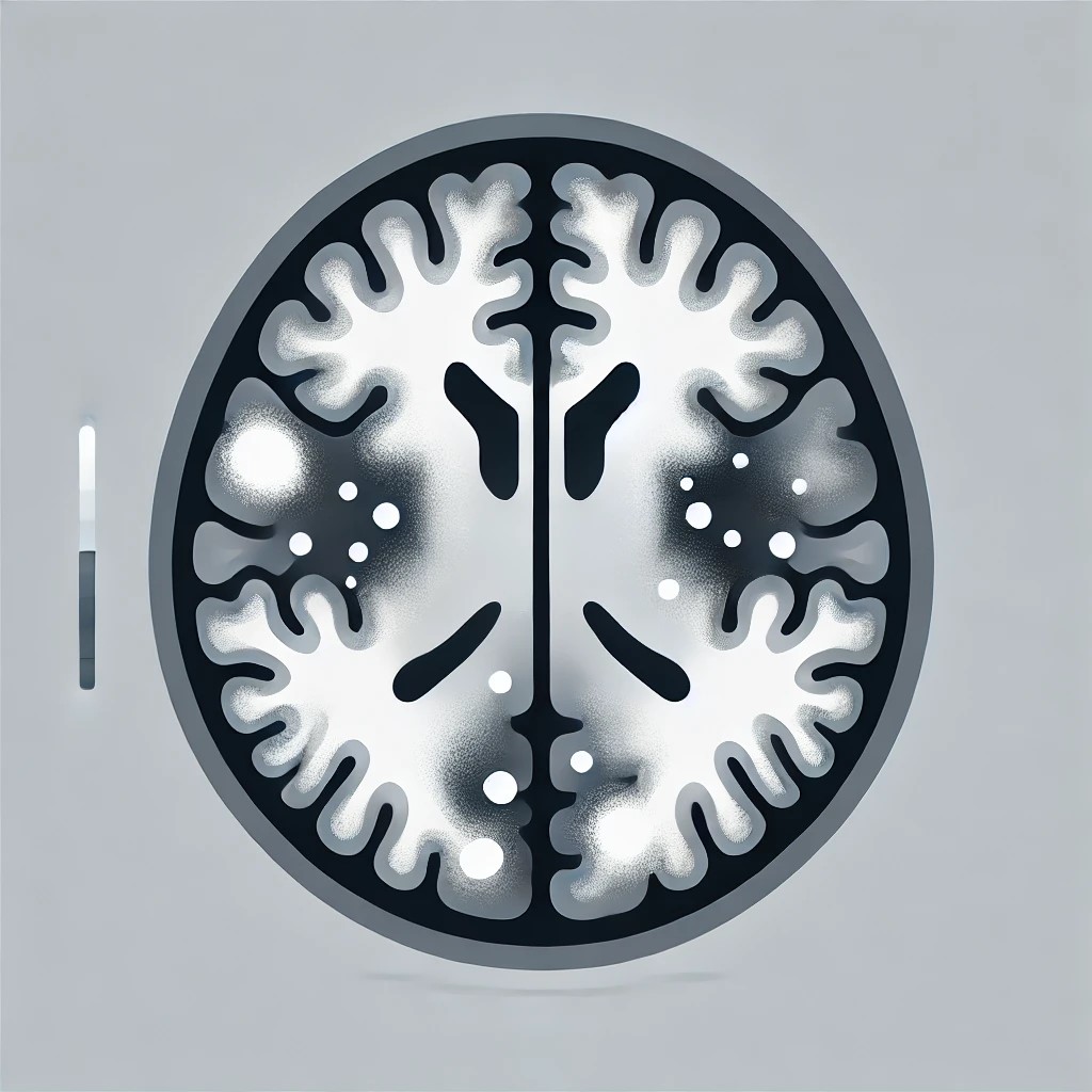
To systematically analyze MRIs for leukoencephalopathies, it is essential to perform a step-by-step evaluation and incorporate serial imaging where necessary. This process involves evaluating white matter involvement, myelination patterns, distinctive imaging characteristics, and integrating clinical presentation for comprehensive diagnosis.
Step 1: Confirm Symmetric White Matter Involvement
- Objective: Identify symmetric white matter abnormalities typical in leukoencephalopathies.
- Method: T2-weighted and FLAIR MRI sequences are essential for detecting and characterizing white matter involvement. While symmetry is typical, exceptions may exist depending on the specific disorder.
Step 2: Determine Hypomyelination or Demyelination
- Objective: Differentiate between hypomyelination (reduced myelin development) and demyelination (loss of previously formed myelin).
- Method:
- Hypomyelination: Appears as persistent T2 hyperintensity with near-normal T1 signal; requires consistent findings on serial MRI, spaced by at least six months after one year of age.
- Demyelination: Characterized by pronounced T2 hyperintensity with T1 hypointensity, indicating myelin breakdown.
Step 3: Assess White Matter Involvement Patterns
- Objective: Classify the white matter involvement pattern to guide potential diagnoses.
- Common Patterns:
- Periventricular: Surrounding the ventricles, seen in multiple sclerosis and leukodystrophies.
- Subcortical: Near cortical areas, associated with leukodystrophies affecting these regions.
- Frontal or Posterior Predominant: Observed in some genetic leukoencephalopathies (e.g., frontotemporal dementia, X-linked adrenoleukodystrophy).
- Diffuse Confluent or Tigroid: Seen in adult-onset autosomal dominant leukodystrophy and metachromatic leukodystrophy.
- Brainstem/Cerebellar Involvement: Specific to some leukoencephalopathies affecting motor control and coordination.
Step 4: Identify Distinctive MRI Features
- Objective: Narrow down differential diagnoses based on specific imaging characteristics.
- Key Features:
- Contrast Enhancement: Suggests inflammation or blood-brain barrier disruption, seen in certain inflammatory or genetic conditions.
- CSF-like Signal Intensity Lesions: Indicates cystic or extensive tissue destruction (e.g., vanishing white matter disease).
- SWI Abnormalities: Reveals microbleeds or calcifications, helpful in vascular or calcific leukoencephalopathies.
- MR Spectroscopy Peaks: Detects abnormal metabolites like lactate, indicating metabolic disorders.
- Spinal Cord Involvement: May indicate longitudinal white matter lesions associated with spinal cord disorders (e.g., LBSL).
Step 5: Correlate MRI with Clinical Presentation
- Objective: Use MRI findings alongside patient age, symptom onset, and progression to refine diagnosis.
- Key Clinical Factors: Motor skill development, neurological signs (e.g., cognitive decline, ataxia), and age at symptom onset can guide the diagnosis, as specific leukodystrophies often present at particular ages or stages.
Step 6: Consider Extra-neurological Manifestations
- Objective: Identify signs outside the nervous system, which can support specific leukodystrophy diagnoses.
- Notable Findings: Adrenal insufficiency, ichthyosis, brittle hair, and photosensitivity can indicate specific disorders like adrenoleukodystrophy or certain metabolic conditions.
Step 7: Employ Ancillary Testing for Confirmation
- Biochemical Testing: Conduct targeted tests (e.g., very long-chain fatty acids, lysosomal enzyme assays) based on MRI findings and suspected disorders.
- Genetic Testing:
- Single-gene testing may be appropriate for specific, strongly suspected conditions.
- Next-generation sequencing or whole-exome/genome sequencing provides a broader diagnostic scope, particularly when initial tests are inconclusive.
Following these systematic steps for MRI analysis, combined with clinical and ancillary diagnostic inputs, enables clinicians to develop a targeted approach for diagnosing leukoencephalopathies, ensuring precise identification, management, and planning.